Breaking news and analysis on politics, business, world national news, entertainment and more.
18+ Homer Wright Rosettes Usmle Pictures
09/02/2015 00:00
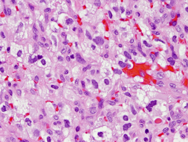
18+ Homer Wright Rosettes Usmle Pictures. Последние твиты от homer wright rosette (@h_wright_rosset). Two rosettes can be seen in this image along with foci of calcification.
Flashcards Table On Neuro Pathology 2 from neuropathology-web.org
Although the cellular mechanisms responsible for the formation of. Popular study materials from usmle step 1. Homer wright rosettes are a type of rosette in which differentiated tumor cells surround the neuropil.
Homer wright rosette — a circular or spherical grouping of dark tumor cells around a pale, eosinophilic, central area that contains neurofibrils but lacks a lumen;
Neuroblastoma, homer wright rosettes are circular groupings of dark tumor cells surrounding pale neurofibrils (small blue cell tumors from neural crest ectoderm). High quality pathology images of endocrine, adrenal, neuroblastoma. Pathology examples of tumors where these can be seen include: It's more of an appendix, it includes images, diagrams and tables of everything you need to know for your 18.